
Dental Radiography, which is among the most important branches in dentistry, holds a significant place in the processes of diagnosis, treatment, and post-treatment evaluation. In recent years, many dentists have replaced old conventional radiography systems with digital imaging systems.
With these systems, digital images created with less radiation exposure to patients can be obtained in a shorter time compared to traditional X-ray films. These digital images can be enhanced, stored, and easily transferred using computer software. Modern digital tomography systems, which are fundamental to today’s high standards, make patient education easier and positively impact communication between patients and dentists.
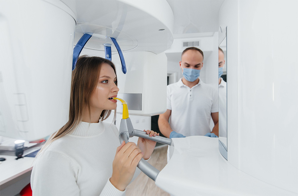
In our clinic, we use the Digital Tomography method in accordance with modern dental standards. So, what are the areas of application of this method that bring our examination and treatment practices to the highest efficiency levels? What kind of conveniences does it provide?
Dental Cone Beam Computed Tomography (CBCT)
Produces detailed and three-dimensional images of the jawbones and teeth, allowing for accurate evaluation of bone volume and width, implant planning, and the localization of impacted teeth.
CBCT also aids in the clear visualization of lesions like cysts within the jawbones. Compared to standard CT scans, CBCT machines emit lower radiation, making them more suitable for patients’ overall health.
CBCT significantly reduces the margin of error before major surgical operations.
Areas of Application: The Dental Tomography method finds application across various phases of diagnosis, treatment, and follow-up procedures, offering substantial benefits to both patients and dentists. The utilization areas and advantages of Dental Tomography can be elucidated as follows:
Detection of Cavities
Prior to and during root canal treatment
Detection of bone damage in cases of advanced gum diseases
Planning and monitoring of implant surgery
Determining the causes of joint disorders
Identifying cysts and tumor suspicions related to teeth and bone
Preoperative determination of impacted tooth positions
Tracking children’s dental development and growth
Clarifying suspicions of dental and jaw fractures
Efficient and rapid solutions for various issues like temporomandibular joint (TMJ) disorders
DENTAL TOMOGRAPHY METHODS
Digital Radiography (RVG)
Digital Radiography utilizes a specialized imaging sensor and a portable radiography device. The image of the patient’s teeth is simultaneously displayed on the computer screen during the procedure. As it eliminates waiting time, it is a practical and fast imaging system. It allows for rapid and detailed imaging of teeth.
Digital Radiography is used for diagnosing interface cavities that are not visible through visual inspection, visualizing initial periapical lesions during root canal treatment, and determining root length during the canal treatment phase.
